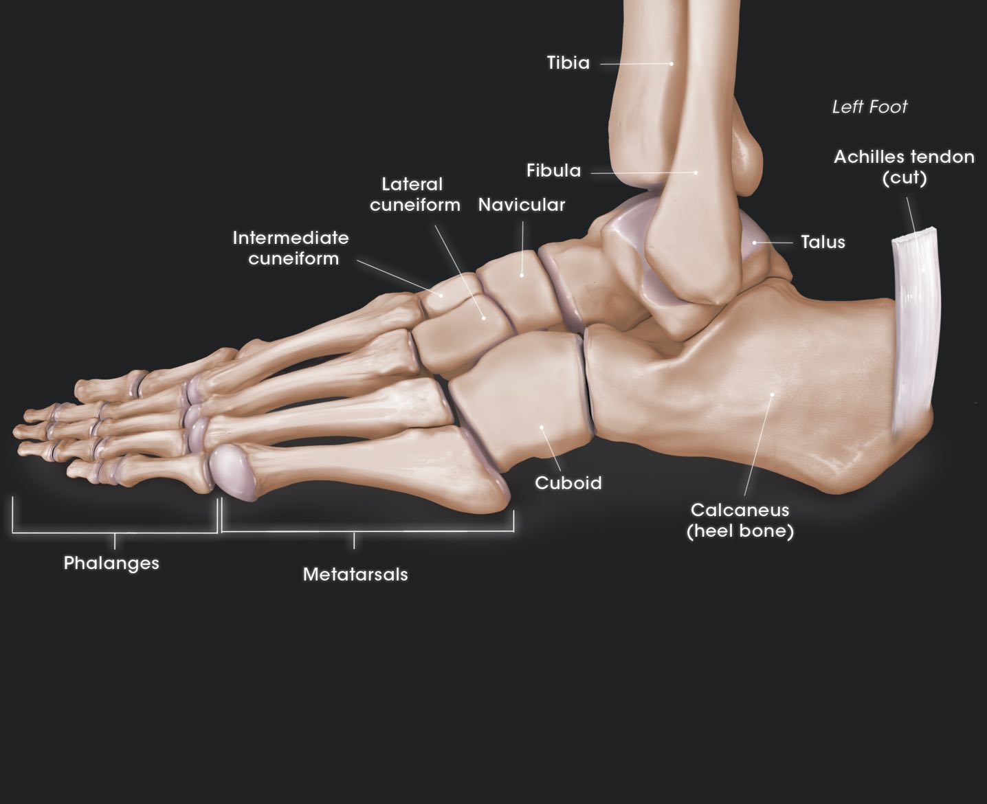
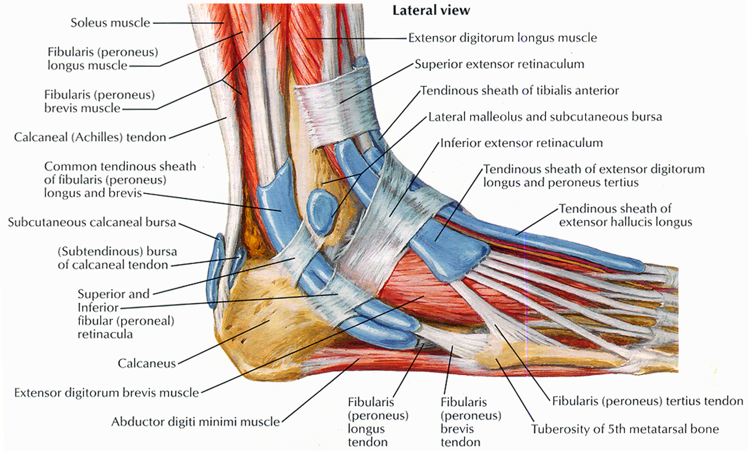
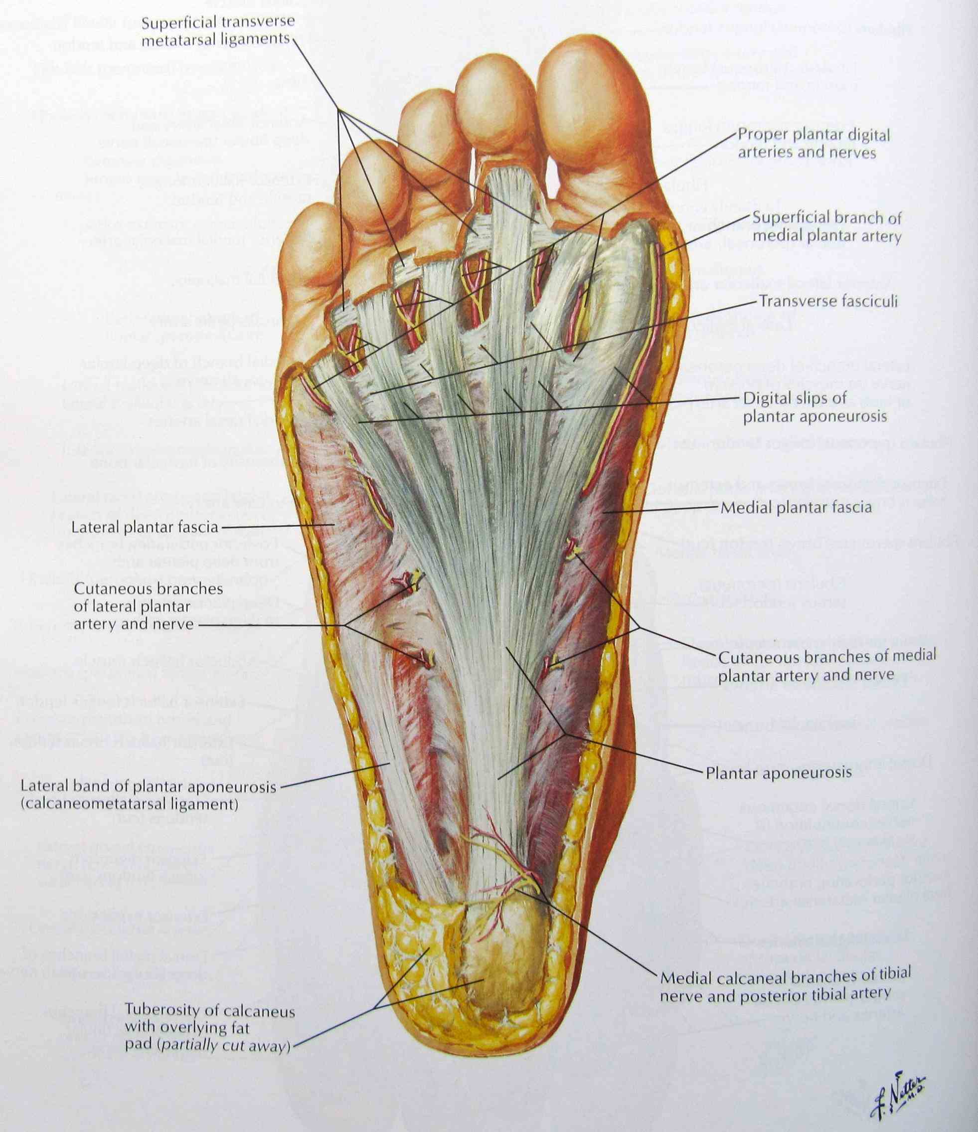
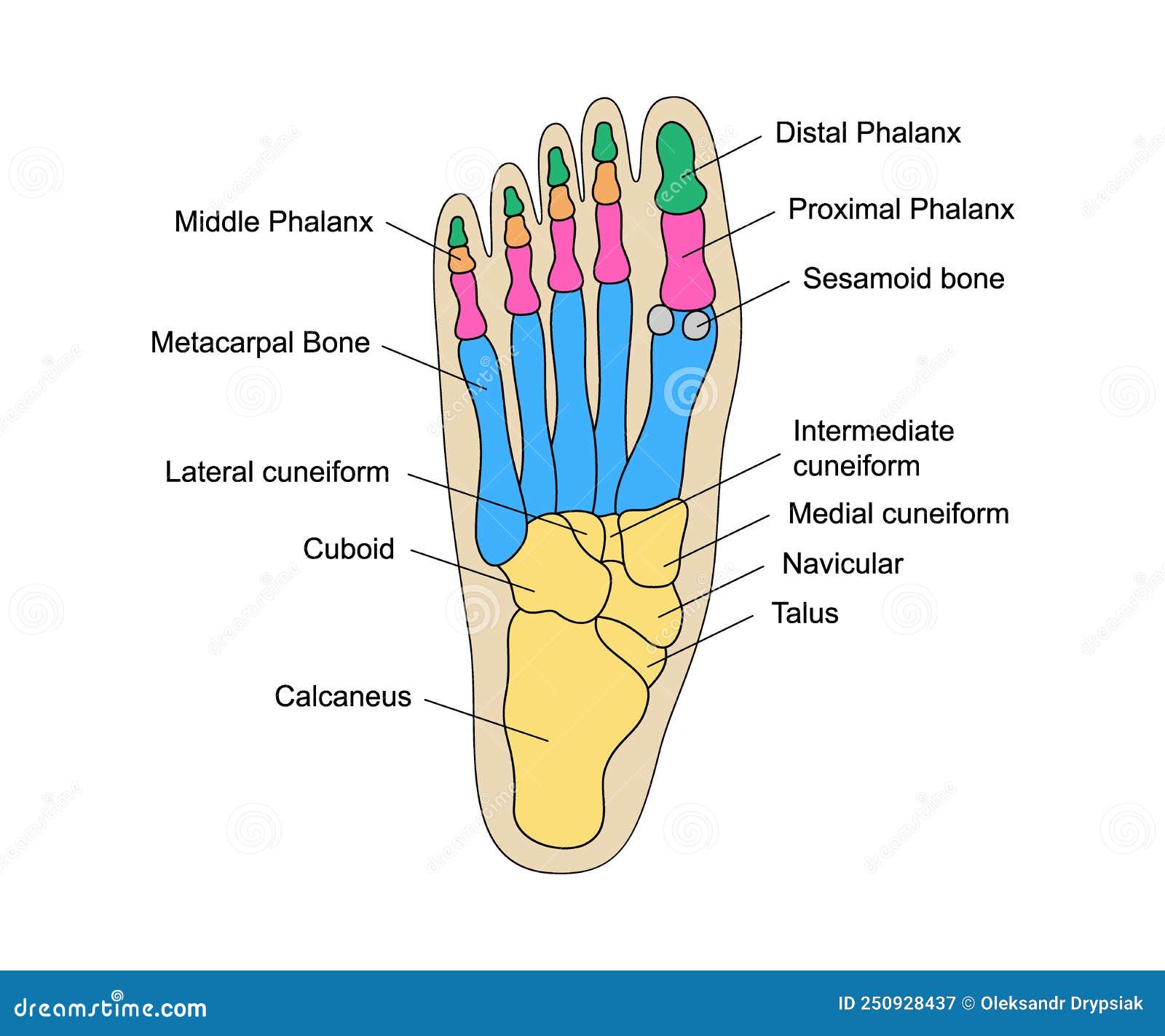
Ligaments are fibrous strands that connect bones. Nerves travel throughout the foot, providing feeling. Nails protect the tips of the toes. Phalanges are the toe bones. Metatarsals are the bones between the toes and the ankle bones. Tarsals are bones of the rear foot (hindfoot) or middle foot (midfoot). The talus is one of the ankle bones.. The Toes, Arch and Heel. Toes are the parts of the foot that allow people to move. They help people grip the ground and push off when they walk or run. The arch is the part of the foot that helps to absorb shock when we move around. It is located between the heel and the toes. The heel provides balance and stability.

Chart of FOOT Dorsal view with parts name Vector image Stock Vector Image & Art Alamy
Patient Education in Orthopedic Surgery OrthoIllustrated

Foot Bones Diagram Quizlet
Foot Anatomy Bones, Muscles, Tendons & Ligaments

Foot Anatomy 101 A Quick Lesson From a New Hampshire Podiatrist Nagy Footcare
Anatomy The Bones Of The Foot
.jpg)
Foot Bones Labeled

Diagram of The Foot 101 Diagrams

Bones of the Lower Limb Anatomy and Physiology
Human Foot Bones Anatomy with Descriptions. Educational Diagram of Internal Organ Illustration

Foot Description, Drawings, Bones, & Facts Britannica
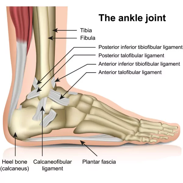
The Basics of Ankle Anatomy and Foot Anatomy
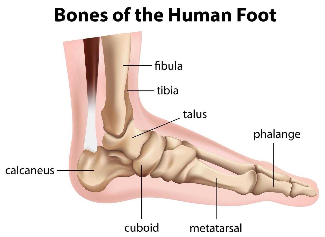
Bones of the human foot diagram 1142236 Vector Art at Vecteezy
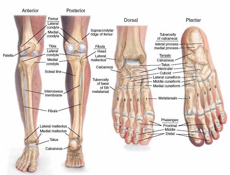
Structure of skeleton of the foot, Tarsals, Metatarsals and Phalanges Science online

Foot & Ankle Bones

Foot and Ankle Anatomical Chart Anatomy Models and Anatomical Charts
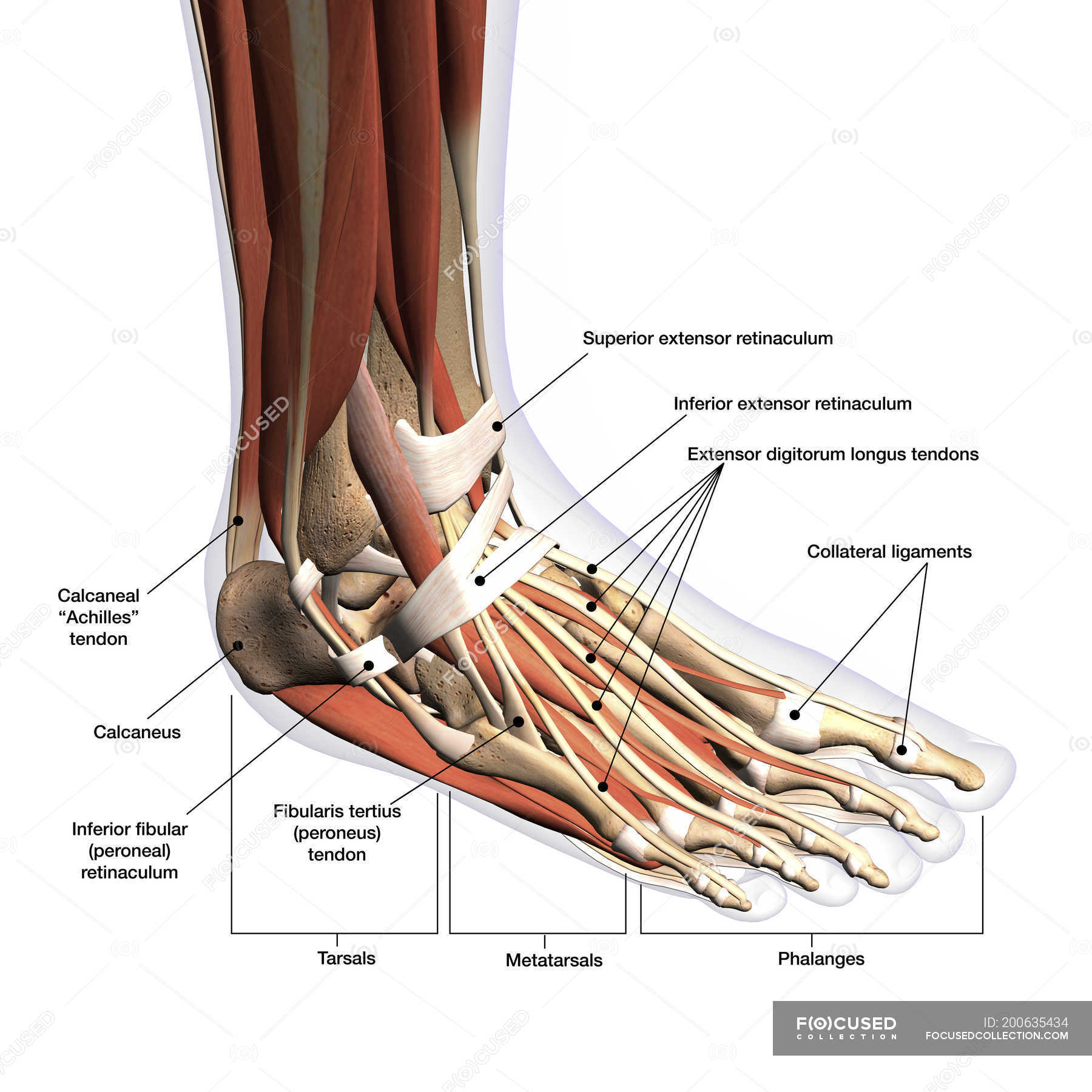
Anatomy of human foot with labels on white background — ankle, leg Stock Photo 200635434

Foot Description, Drawings, Bones, & Facts Britannica

Anatomy of the Foot Comprehensive Orthopaedics
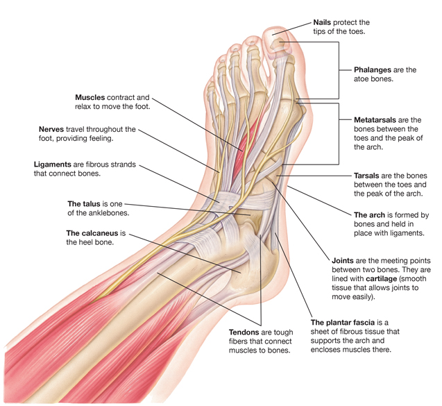
Parts of a Foot
The midfoot is a pyramid-like collection of bones that form the arches of the feet. These include the three cuneiform bones, the cuboid bone, and the navicular bone. The hind foot forms the heel and ankle. The talus bone supports the leg bones (tibia and fibula), forming the ankle. The calcaneus (heel bone) is the largest bone in the foot.. Ankle anatomy. The ankle joint, also known as the talocrural joint, allows dorsiflexion and plantar flexion of the foot. It is made up of three joints: upper ankle joint (tibiotarsal), talocalcaneonavicular, and subtalar joints.The last two together are called the lower ankle joint. The upper ankle joint is formed by the inferior surfaces of tibia and fibula, and the superior surface of talus.